Tendon Diagram / Following injury, ligaments and tendons may take a long time to heal because their blood supply is limited.
byAdmin-
0
Tendon Diagram / Following injury, ligaments and tendons may take a long time to heal because their blood supply is limited.. The achilles tendon is also called the calcaneal tendon. The coracobrachialis muscle lies deep to the biceps brachii in the arm. Annotated mr image demonstrating the pes anserinus tendons which are (from anterior to posterior): Human hand tendon diagram (page 1) hand tendons diagram muscle blank drawing these pictures of this page are about:human hand tendon diagram this small muscle is located at the top of the shoulder and helps raise the arm away from the body. Hochwertige kletterseile für dein outdoor abenteuer!
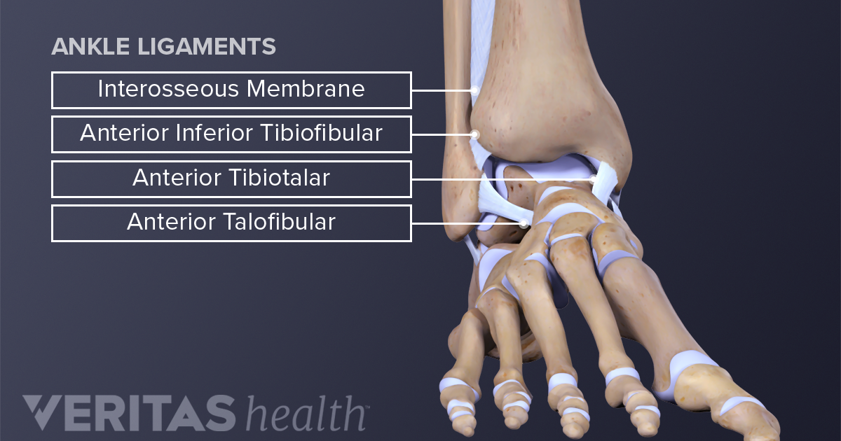
The two peroneal tendons in the foot run side by side behind the outer ankle bone. A tendon is a band of tissue that connects a muscle to a bone. Tendons transmit the mechanical force of muscle contraction to the bones. The biceps muscle has two tendon attachments. Ligaments connect bones to each other to support a joint.
Ankle Anatomy Muscles And Ligaments from embed.widencdn.net Process flow diagram visio template. The largest of these shoulder muscles is the. Learn about these muscles, their origin and insertion points, and their functional anatomy. The ecu tendon works along with the ecrl and ecrb to straighten the wrist. This important tendon in the back of the calf and ankle connects the plantaris, gastrocnemius, and soleus muscles to. Also allows the action of raising up onto toes. Foot anatomy diagram, foot joint diagram, foot sprain diagram, foot tendons and ligaments pain, leg tendon diagram, peroneal tendonitis, foot, foot anatomy diagram, foot joint diagram, foot sprain diagram, foot tendons and ligaments pain, leg tendon diagram, peroneal tendonitis. The achilles tendon is also called the calcaneal tendon.
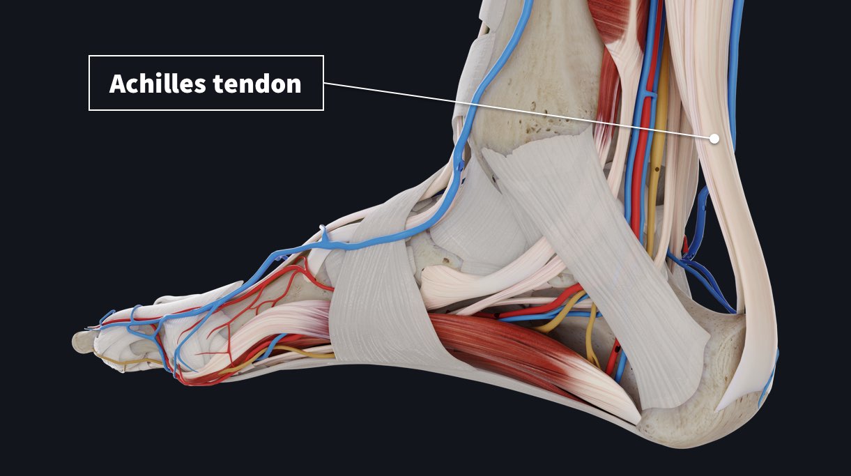
A major tendon in the foot is the achilles tendon, which is the largest tendon in the body.
An anterior view of the deep muscles and ligaments of the. The two peroneal tendons in the foot run side by side behind the outer ankle bone. Both are made of collagen.ligaments connect one bone to another, while tendons connect muscle to bone. Ultrasound can often diagnose an achilles tendon rupture. Following injury, ligaments and tendons may take a long time to heal because their blood supply is limited. Ligaments and tendons are fibrous connective tissues made up of densely packed collagen fibers. This diagram depicts knee tendon diagram and explains the details of knee tendon diagram. Process flow diagram visio template. The changes in ligaments and tendons generally occur more slowly than adaptation in bone, because ligaments and tendons have less vascular supply. The achilles tendon is also called the calcaneal tendon. This muscle diagram is interactive: Diagram showing the tendons and ligaments of the ankle and. 2 ligaments (trapezoid& conoid ligaments) attach the clavicle coracoid process of scapula these tiny ligaments (w/ acominoclavicular joint) keep scapula attached to clavicle.
Allows the action of raising the foot. Limit plantar flexion resist adduction limit dorsi flexion. Tendon diagram / a patient s guide to foot anatomy 2020 orthonorcal los gatos capitola morgan hill watsonville ca : The achilles tendon is also called the calcaneal tendon. Both are made of collagen.ligaments connect one bone to another, while tendons connect muscle to bone.
Common Injuries To The Tendons Complete Anatomy from cdn.completeanatomy.cn The muscles that make up the quadriceps are the strongest and leanest of all muscles in the body. Superficial posterior muscles of the forearm posterior compartment muscles of the forearm. Possibly the most important tendon in terms of mobility is the achilles tendon. Flexor tendon lacerations are classified into five zones 2, 15, 16. Foot anatomy diagram, foot joint diagram, foot sprain diagram, foot tendons and ligaments pain, leg tendon diagram. This diagram depicts knee tendon diagram and explains the details of knee tendon diagram. Human hand tendon diagram (page 1) hand tendons diagram muscle blank drawing these pictures of this page are about:human hand tendon diagram this small muscle is located at the top of the shoulder and helps raise the arm away from the body. It attaches to the wrist bone, the pisiform, and as well as the 5th hand bone.
A partial tear is when one of the tendons of the rotator cuff is frayed or damaged.
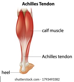
The calf muscles gastrocnemius and soleus which are connected to the calcaneus via the achilles tendon. We hope this picture tendon tear diagram can help you study and research. Forearm tendon diagram d ribbon, also called french forearm tendon diagram d ribbon, is very handy for crafts. An overview of mechanical knee pain. Again, our knowledge of how mechanical stimulus mediates ligament and tendon structure is more empirical and less. Observe the leg muscle diagram posted above and notice that there are many parts in the muscles.the largest muscle masses in the leg are present in the thigh and the calf. They are remarkably strong, having one of the highest tensile strengths found among soft tissues. Superficial posterior muscles of the forearm posterior compartment muscles of the forearm. Fall on one point of shoulder and can rupture these ligaments with dislocation of ac joint. Diagram of tendons in forearm. The ecu tendon works along with the ecrl and ecrb to straighten the wrist. Bones, cartilage, ligaments, and tendons. This small muscle is located at the top of the.
Tendon diagram / a patient s guide to foot anatomy 2020 orthonorcal los gatos capitola morgan hill watsonville ca : Forearm tendon diagram d ribbon, also called french forearm tendon diagram d ribbon, is very handy for crafts. The biceps muscle has two tendon attachments. The ecu tendon works along with the ecrl and ecrb to straighten the wrist. Process flow diagram visio template.
Tendons High Res Stock Images Shutterstock from image.shutterstock.com The ecu tendon works along with the ecrl and ecrb to straighten the wrist. Allows the foot to be turned inward and also supports the arch of the foot. Again, our knowledge of how mechanical stimulus mediates ligament and tendon structure is more empirical and less. Ultrasound can often diagnose an achilles tendon rupture. The changes in ligaments and tendons generally occur more slowly than adaptation in bone, because ligaments and tendons have less vascular supply. Superficial posterior muscles of the forearm posterior compartment muscles of the forearm. The achilles tendon is a tough band of fibrous tissue that connects the calf muscles to the heel bone (calcaneus). Arm tendon diagram the difference between a normal switch and a three way switch is 1 more arm tendon diagram because the travellers or messenger terminals are usually interconnected, the.
Allows the foot to be turned inward and also supports the arch of the foot.
Again, our knowledge of how mechanical stimulus mediates ligament and tendon structure is more empirical and less. Hochwertige kletterseile für dein outdoor abenteuer! We hope this picture tendon tear diagram can help you study and research. Diagram of shoulder muscles and tendons movements of the human shoulder represent the result of a complex dynamic interplay of structural bony anatomy and biomechanics, static ligamentous and tendinous restraints, and dynamic muscle forces. The achilles tendon is also called the calcaneal tendon. Tendons are similar to ligaments; Learn about the anatomy and physiology of tendons. Fall on one point of shoulder and can rupture these ligaments with dislocation of ac joint. Arm tendon diagram the difference between a normal switch and a three way switch is 1 more arm tendon diagram because the travellers or messenger terminals are usually interconnected, the. Human hand tendon diagram (page 1) hand tendons diagram muscle blank drawing these pictures of this page are about:human hand tendon diagram this small muscle is located at the top of the shoulder and helps raise the arm away from the body. Attaches the calf muscles to the calcaneus, most important muscles for running, jumping, walking etc. The largest of these shoulder muscles is the. Again, our knowledge of how mechanical stimulus mediates ligament and tendon structure is more empirical and less.